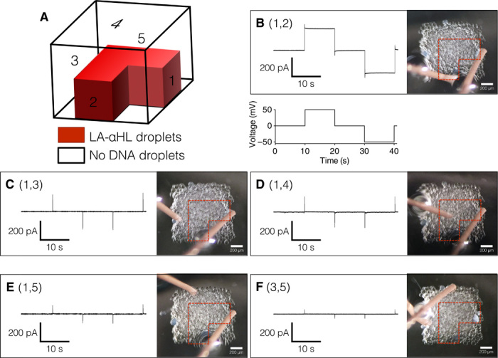
Fig. 7. Electrical recordings from an L-shaped pathway formed by expression from LA-αHL DNA in a 3D-printed synthetic tissue.

(A) Schematic of the printed tissue containing droplets with LA-αHL DNA (red) printed with droplets containing no DNA (clear region within black frame). Numbers represent sides of the cuboid where electrodes were placed to detect the conductive pathway. (B to F) Electrical recordings detect a current when the electrodes are at positions 1 and 2 (B), based on the voltage protocol in (B), but not when one or both of the electrodes are positioned off the pathway sides 1 and 3 (C), 1 and 4 (D), 1 and 5 (E), or 3 and 5 (F).
