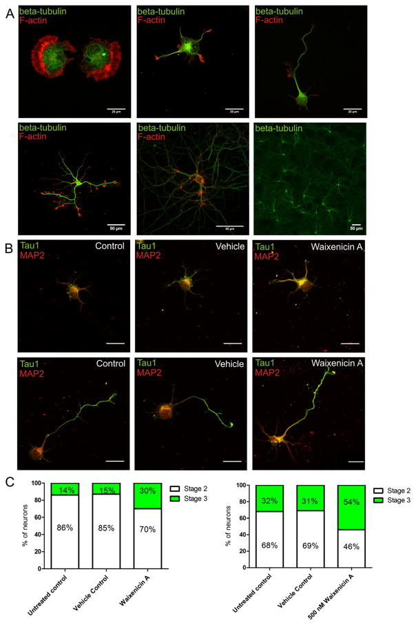
Fig. 5.
Treatment with waixenicin A promotes maturation of hippocampal neurons. a Representative images of developmental stages of hippocampal neurons according to Dotti et al. Top panel: stage 1 (left), stage 2 (center), and stage 3 (right). Bottom panel: stage 4 (left) and stage 5 (center and right). b Representative images of neurons in stage 2 (top panel) and stage 3 (bottom panel) in different treatment groups. Scale bars are 20 μm. c Proportion of neurons in stages 2 and 3 in different treatment groups. Left panel: DIV2 cultures (6 h post-treatment; n=137); right panel: DIV3 cultures (24 h post-treatment; n=144). Chi-squared: DIV2 untreated control versus waixenicin A, p=0.006; DIV3 untreated control versus waixenicin A, p=0.04

