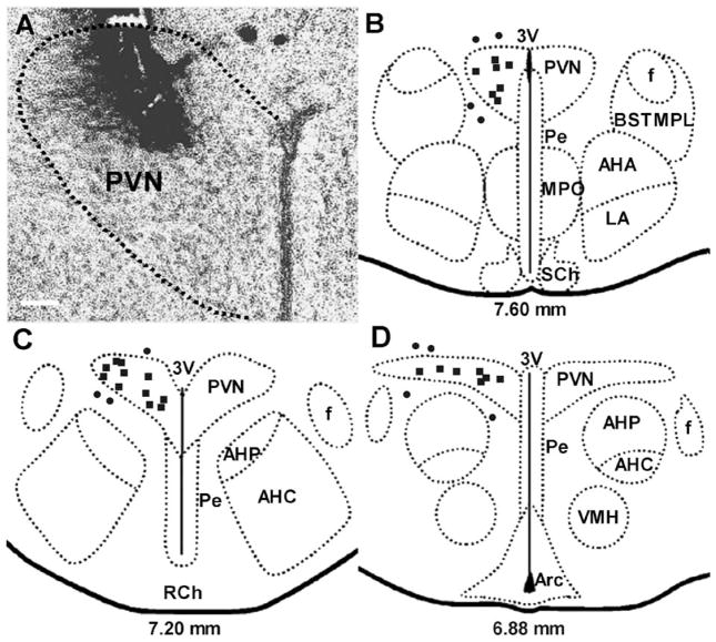
Fig. 1.
(A) Light-field microphotograph showing the site of termination of an injection that is considered to be within the PVN. (B, C and D) Schematic representations of serial sections from 7.60 to 6.88 mm anterior to interaural zero showing the sites of termination of injection tracts in 36 rats. Each square represents a site of termination of an injection that is considered to be within the PVN, while each circle represents a site of termination of an injection that is considered to be outside of the PVN. The scale bar in (A) = 0.1 mm. 3V, third ventricle; AHA, anterior hypothalamic area, anterior part; AHC, anterior hypothalamic area, central part; AHP, anterior hypothalamic area, posterior part; Arc, arcuate hypothalamic nucleus; BSTMPL, bed nucleus of the stria terminalis, medial division, posterolateral part; f, fornix; LA, lateroanterior hypothalamic nucleus; MPO, medial preoptic nucleus; Pe, periventricular hypothalamic nucleus; PVN, paraventricular nucleus; RCh, retrochiasmatic area; VMH, ventromedial hypothalamic nucleus. Drawing is modified from Paxinos and Watson [35].

