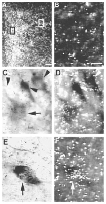
Fig. 7.
(A) Dark-field photomicrograph of the PVN showing localization of RAMP-2 mRNA with in situ hybridization. (B) Dark-field photomicrograph showing the background signal in an area near the PVN. (C and D) Light-and dark-field photomicrographs of the parvocellular subdivision at higher magnification, indicated by the black box in (A) and showing single-labeled neurons (arrowhead) and a neuron double-labeled with RAMP-2 mRNA and NADPH-d (arrow). (E and F) Light- and dark-field photomicrographs of the magnocellular subdivision at higher magnification, indicated by the white box in (A) and showing a neuron double-labeled with RAMP-2 mRNA and NADPH-d (arrow). The scale bar in (A) = 100 μm; the scale bar in (B) = 10 μm and applies in (C, D, E and F). V, third ventricle.

