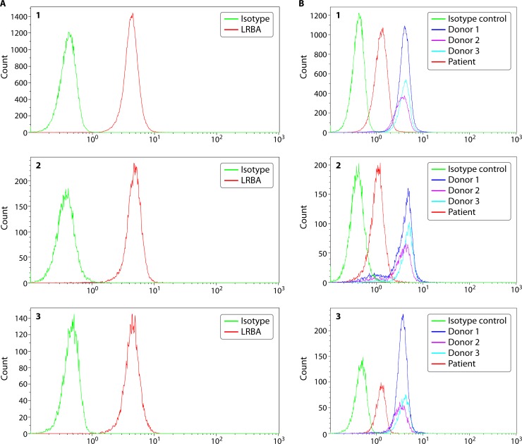
FIG 2.
LRBA protein expression in lymphocytes from a healthy individual and a patient. LRBA protein is expressed intracellularly, and data are shown for T cells (panel 1), B cells (panel 2), and NK cells (panel 3) from a healthy donor (A) and a patient (B). Peripheral blood mononuclear cells (PBMCs) are isolated from blood samples collected in sodium heparin or EDTA and assessed for LRBA expression without stimulation, using an isotype control and a specific primary antibody. Intracellular protein expression is assessed by cell fixation and permeabilization prior to simultaneous staining with cell lineage markers and primary antibody. The protein is visualized using a fluorescently labeled secondary antibody. Both percent-positive lymphocyte subsets (T, B, or NK cells) along with mean fluorescence intensity (MFI) information are captured. LRBA protein is robustly expressed in the majority of lymphocyte subsets without stimulation. In the B panels, data from three healthy donors are represented as donor 1, donor 2, and donor 3. The patient shows a normal proportion of lymphocyte subsets expressing LRBA; however, the mean fluorescence intensity, which correlates with the amount of protein expression, is significantly reduced, which is consistent with LRBA deficiency. The patient had a clinical phenotype of very early onset inflammatory bowel disease, failure to thrive, and multiple autoimmune manifestations. A homozygous 2-bp deletion was identified (c.3958_3986del; p.D1329Yfs*18) in the LRBA gene.

