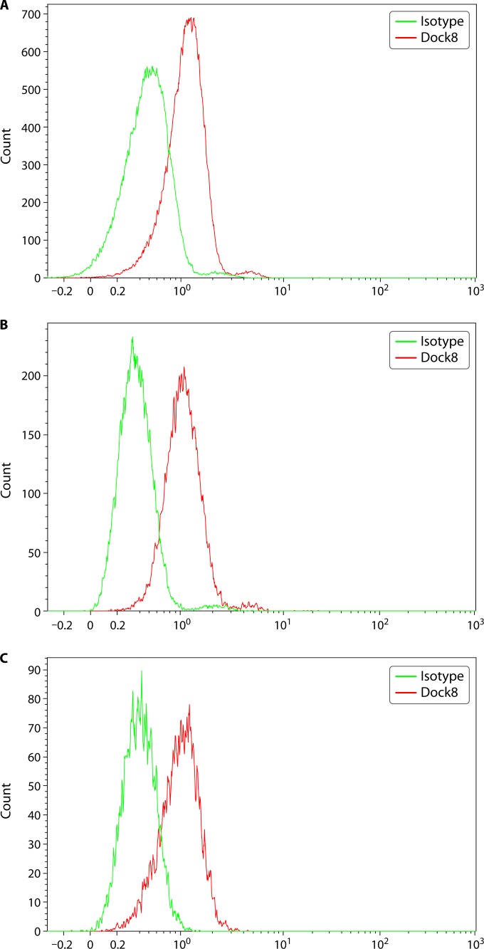
FIG 3.
DOCK8 protein expression in lymphocytes from a healthy individual. DOCK8 protein is expressed intracellularly, and data are shown for T cells (A), B cells (B), and NK cells (C). Peripheral blood mononuclear cells (PBMCs) are isolated from blood samples treated with sodium heparin or EDTA and assessed for DOCK8 expression without stimulation, using an isotype control and a specific primary antibody. Intracellular protein expression is assessed by cell fixation and permeabilization prior to simultaneous staining with cell lineage markers and primary antibody. The protein is visualized using a fluorescently labeled secondary antibody. Both percent-positive lymphocyte subsets (T, B, or NK cells) along with mean fluorescence intensity (MFI) information are captured. DOCK8 protein is robustly expressed in the majority of lymphocyte subsets without stimulation.

