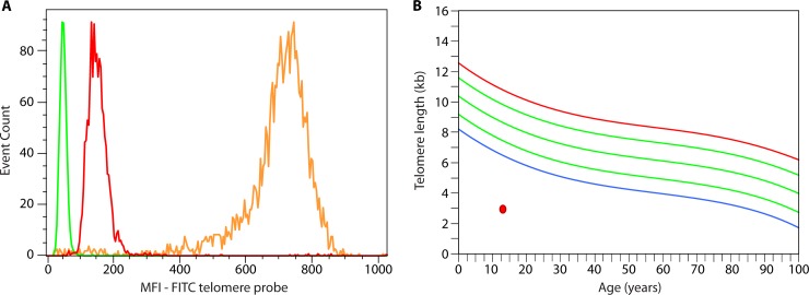
FIG 6.
Assessment of lymphocyte telomere length in a patient with dyskeratosis congenita. (A) Leukocyte telomere length assessment is performed by measuring the median fluorescence intensity (MFI) of distinct cell subpopulations while controlling for DNA content (not shown) and controlling for the intrinsic fluorescent properties of the cell type analyzed (green [performed in a separate tube and overlaid on the graph]). The MFI of the gated lymphocyte cell population is shown on the graph (red), along with the internal positive hybridization control cells (fixed cow thymocytes [orange]). FITC, fluorescein isothiocyanate. (B) An example of a young patient with dyskeratosis congenita showing in the selected lymphocyte cell population example very short lymphocyte telomere length (red circle) compared to the reference curve summarizing data from healthy individuals – or below the first percentile of distribution for age (blue curve); green curves represent the 10th, 50th, and 90th percentiles, and the red curve represents the 99th percentile of distribution.

