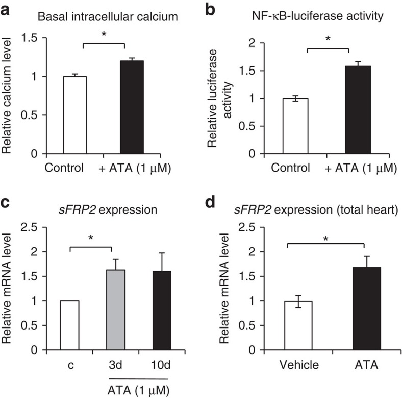
Figure 6. PMCA4 inhibition increased sFRP2 levels in cardiac fibroblasts and the heart.
(a) ACFs isolated from WT mice were subjected to ATA treatment (1 μM for 48 h). Measurement of basal intracellular calcium using fluo-3 dye indicated a significantly higher calcium level in ATA-treated cells (n=4; *P<0.05, Student's t-test). (b) Analysis of NF-κB activity using the NF-κB-luciferase construct showed a significant increase in NF-κB activity in ATA-treated cells (n=4; *P<0.05, Student's t-test). (c) ACFs were treated with ATA (1 μM) for 3 or 10 days. qRT–PCR analysis showed that sFRP2 level was significantly enhanced in ATA-treated cells (n=4; *P<0.05, Student's t-test). (d) WT mice were injected with ATA (5 mg kg−1 per day for 2 weeks). sFRP2 level was detected in total heart's mRNA. Results showed a significant increase in sFRP2 level (n=6–8 in each group; *P<0.05, Student's t-test). All error bars represent the s.e.m.

