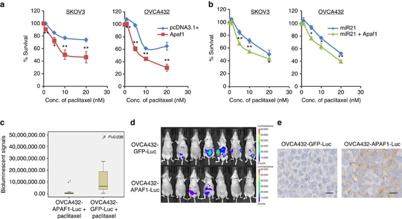
Figure 6. APAF1 mediates miR21-induced paclitaxel resistance.
(a) Overexpression of APAF1 by full-length transfection increased paclitaxel sensitivity in ovarian cancer SKOV3 and OVCA432 cells. The results were the average from at least three independent experiments. Mean±s.d.; **P<0.01 and *P<0.05; two-tailed Student's t-test. (b) Co-transfection of miR21 precursor and full-length APAF1 decreased paclitaxel resistance compared with the co-transfection of pre-miR21 and the control vector in both SKOV3 and OVCA432 cells. The results were the average from at least three independent experiments. Mean±s.d.; **P<0.01 and *P<0.05; two-tailed Student's t-test. (c–e) APAF1 overexpression enhanced the paclitaxel sensitivity of ovarian cancer cells in vivo. APAF1 stably overexpressing ovarian cancer OVCA432 cells were generated using the lentiviral transduction method and were intraperitoneally injected into female BALB/c athymic nude mice, followed by paclitaxel treatment. The tumour volumes were measured and quantified using the IVIS-Lumina XR in vivo imaging system after a 2-week 5 mg kg−1 paclitaxel treatment. (c) Box plot showing a significant decrease in luciferase activity in the APAF1 overexpression group (n=7) compared with the control group (n=7) after paclitaxel treatment (P=0.038; Mann–Whitney U-test). (d) Representative images show a decrease in luminescence in the APAF1 overexpression group compared with the control group after paclitaxel treatment. (e) Immunolocalization of APAF1 on paraffinized sections of tumour tissues collected from mice demonstrated a higher APAF1 level in the APAF1 overexpression group (n=7) compared with the control group (n=7). Representative microscopic images were illustrated. Scale bar, 10 μm.

