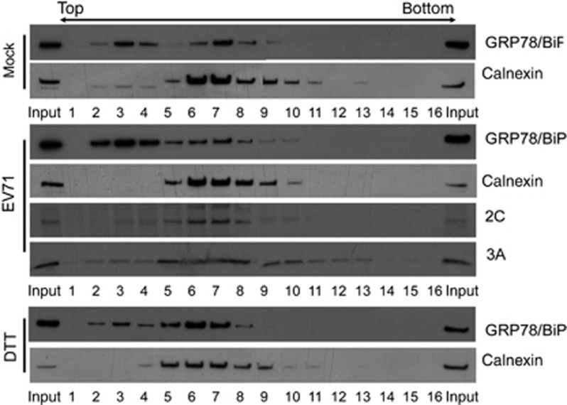
Figure 1.
Subcellular fractionation profiles of GRP78/BiP in mock-infected, EV71-infected or DTT-treated RD cells. The PNSs from mock-infected, EV71-infected (MOI of 10, 6 h p.i.), 2.5 mM DTT-treated (6 h) cells were fractionated using OptiPrep gradients. The fractions were collected from the top and subjected to SDS–PAGE and subsequent western blotting using specific antibodies against GRP78/BiP, calnexin, and viral proteins 3A and 2C. The results are representative of three independent experiments.

