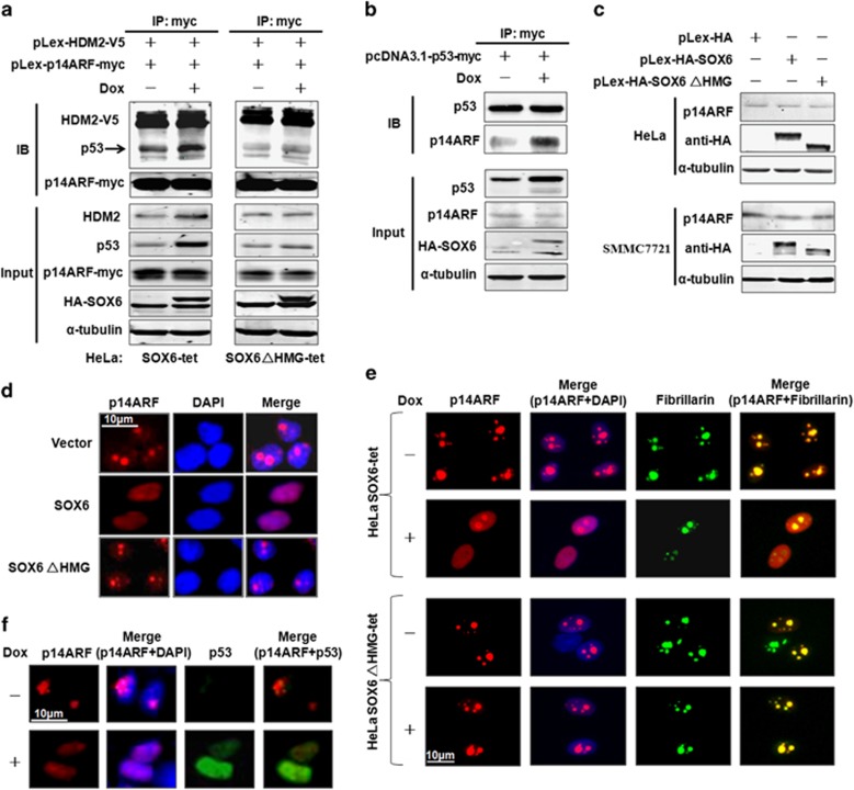
Figure 5.
SOX6 promotes formation of the p14ARF-HDM2-p53 ternary complex and translocation of p14ARF to the nucleoplasm. (a) HeLa SOX6-tet and SOX6ΔHMG-tet cells were co-transfected with pLex-p14ARF-myc or pLex-HDM2-V5 plasmids with or without Dox (2 μg/ml) treatment, and the cell lysates were subjected to co-immunoprecipitation by anti-myc-tag antibody. One-tenth of the total cell lysate volume was subjected to direct western blot analysis. (b) HeLa SOX6-tet cells were transfected with the pCDNA3.1-p53-myc plasmid and the cell lysates were subjected to co-immunoprecipitation (co-IP) by anti-myc-tag antibody. (c) Western blot was performed to analyze the p14ARF protein levels in HeLa and SMMC7721 cells that had been transfected with pLex-HA-SOX6, pLex-HA-SOX6ΔHMG or control expression vector. (d) Immunocytofluorescence staining of p14ARF in HeLa cells that had been co-transfected with pLex-p14AF and pLex-HA-SOX6, pLex-HA-SOX6ΔHMG or control expression vector. (e) Immunocytofluorescence staining of p14ARF in HeLa SOX6-tet and SOX6ΔHMG-tet cells that had been transfected with pLex-p14ARF and treated with or without Dox (2 μg/ml) for 48 h. Fibrillarin was used as a nucleolar marker. (f) Immunocytofluorescence staining of p14ARF and endogenous p53 in HeLa SOX6-tet cells that had been transfected with pLex-p14ARF and treated with or without Dox (2 μg/ml) for 48 h. IP, immunoprecipitate; IB, immunoblotting.

