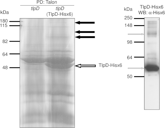
Figure 1. Pull-down of TlpD-Hisx6 with potential protein interaction partners from H. pylori N6 cleared cell lysates.
Pull-down (PD) precipitates of TlpD-Hisx6 from N6 tlpD (TlpD-Hisx6) and N6 tlpD (negative control not expressing any TlpD; precipitated using Talon (Cobalt2+–coupled matrix)) were separated in 12% SDS gels and stained with Coomassie blue (left panel). Black arrows point at specific higher molecular mass protein bands that were detected only in the pull-down material of H. pylori N6 tlpD (TlpD-Hisx6) but not in the N6 tlpD mutant (control strain). These gel sections, containing differentially detected bands which are numbered 1, 2, 3 from top to bottom (corresponding to numbers in Table 1), were subsequently cut from the gels separately and analysed using mass spectrometry (MS/MS; see Supplementary Methods). The white arrow points at TlpD-Hisx6 monomer. The right panel shows a corresponding Western blot (WB) of the purified TlpD-Hisx6 fraction which was developed using α-Hisx6 antibody. The arrows in the right panel point at TlpD-Hisx6 monomer (lower arrow) and TlpD-Hisx6 dimer (upper arrow).

