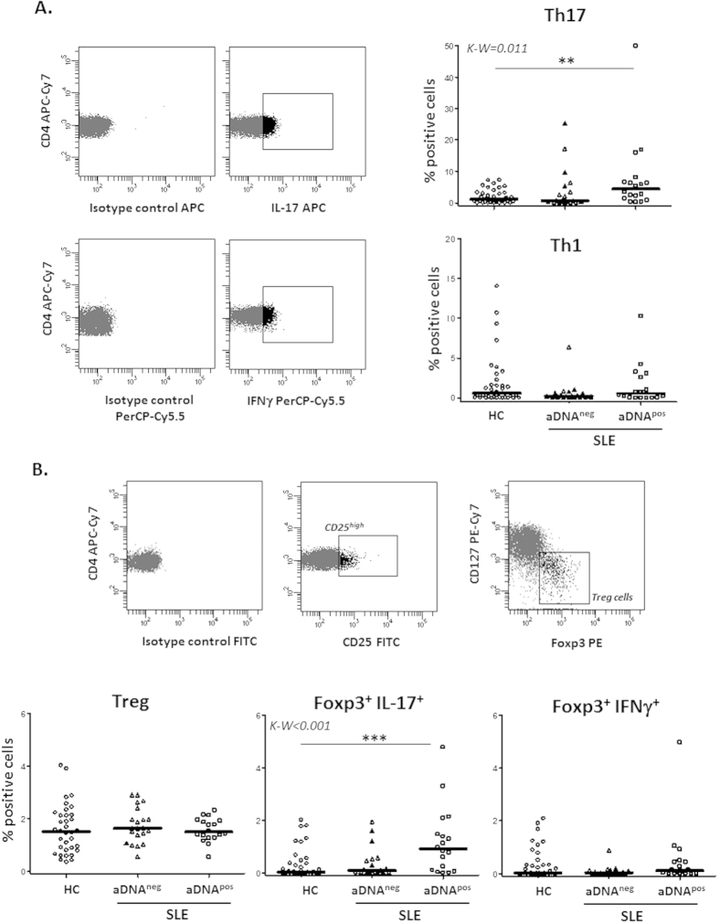
Figure 2. Increased IL-17 producing cells in SLE patients with anti-dsDNA antibodies.
Foxp3, CD25, CD127, IL-17 and IFNγ expression was analyzed in fresh peripheral blood CD4+ lymphocytes from SLE patients and HC. (A) Dot-plots show cells positive for IL-17 or IFNγ expression, determined attending to the fluorescence of cells labelled with the corresponding isotype-matched conjugated irrelevant MAb as a negative control. Scatter plots represent the percentage of IL-17+ (Th17) and IFNγ+ (Th1) CD4+ cells in HC and SLE patients presenting (pos) or not (neg) anti-dsDNA antibodies (aDNA). Horizontal bars show the median. (B) Treg cells were sequentially identified as CD4+CD25highCD127lowFoxp3+ cells. Scatter plots represent the quantity of Treg, Foxp3+ IL-17+ and Foxp3+ IFNγ+ cells in HC and SLE patients in function of their anti-dsDNA status, and horizontal bars show the median. Statistical differences among groups were evaluated by Kruskal-Wallis test and Dunn’s post test was conducted to determine which groups’ pairs had different means. **p < 0.01; ***p < 0.001.

