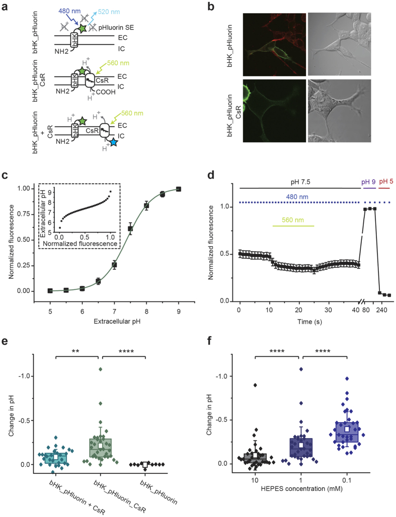
Figure 5. pH changes at the extracellular membrane surface in HEK293 cells.
(a) Protein architectures of bHK_pHluorin (ß-subunit of the HK-ATPase fused to superecliptic pHluorin), bHK_pHluorin_CsR (bHK_pHluorin fused to the N-terminus of CsR T46N) and bHK_pHluorin + CsR (bHK_pHluorin coexpressed with CsR_T46N_eCFP via a P2A cleavage site). (b) Confocal images of bHK_pHluorin and bHK_pHluorin_CsR (pHluorin in green, Rhodamine18 membrane marker in red (only for bHK_pHluorin)). (c) Normalized fluorescence of bHK_pHluorin as a function of extracellular pH (mean +/− SD, n = 9). Inset: Extracellular pH dependence on normalized fluorescence. (d) Experimental protocol for the measurement of extracellular pH changes: bHK_pHluorin_CsR was illuminated for 15 s by 560 nm light with simultaneous fluorescence measurements of pHluorin (480 nm excitation pulses) (mean +/− SEM, n = 25), calibrated by fluorescence measurements at pH 9 and pH 5. (e) Extracellular pH changes after 560 nm light in 1 mM HEPES in absence of a proton pump (bHK_pHluorin, n = 10), in direct vicinity to a proton pump (bHK_pHluorin_CsR, n = 35) and on the overall membrane surface in presence of CsR_T46N, (bHK_pHluorin + CsR, n = 27) (box represents 25% to 75% percentile, empty square represents mean + − SD, two sample t-test with Welch’s correction with t = −3.1l, df = 47.7, p = 0.0032 for the comparison of bHK_pHluorin + CsR to bHK_pHluorin_CsR and t = 5.97, df = 36.17, p < 0.0001 for the comparison of bHK_pHluorin_CsR to bHK_pHluorin). (f) pH changes in direct vicinity to CsR T46N measured with bHK_pHluorin_CsR in different extracellular proton buffer concentrations (n = 35, two-sample paired t-test with t = −6.24, df = 34, p < 0.0001 for the comparison of 10 mM HEPES to 1 mM HEPES and t = −7.03, df = 34, p < 0.0001 for the comparison of 1 mM HEPES to 0.1 mM HEPES). Photocurrents of the proton pump constructs were comparable but variable and overall small reflecting the small pH changes observed with the fluorescent dye (45 +− 30 pA for bHK_pHluorin_CsR and 55 +− 45 pA for the CsR_eCFP_P2A_bHK_pHluorin split construct).

