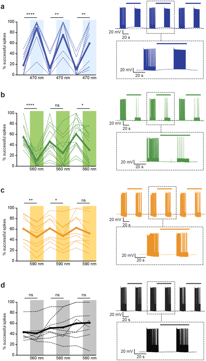
Figure 7. Functional impact of bystander currents in neurons at spike threshold.
To facilitate effect detection, spikes were evoked in the bystander neuron by intracellular injection of electrical current pulses at 10 Hz with current magnitudes titrated to spike threshold, to achieve ~50% spike success rate at baseline. In dense opsin-expressing regions, light stimulation was then applied and the change in evoked spiking of bystander neurons was recorded. Plots show percentage of successfully evoked spikes during repeated light-off and light-on epochs for (a), AAV-ChR2 (RM one-way ANOVA: F = 28.78, n = 10 cells, p < 0.0001). (b) AAV-eArch3.0 (F = 12.16, n = 13 cells, p < 0.0001). (c) AAV-eNpHR3.0 (F = 8.361, n = 9, p = 0.030). (d) AAV-YFP control bystander neurons (F = 2.813, n = 8 cells, p = 0.1197). In summary plots (left), thick lines indicate group mean and thin lines indicate individual cell data. Example traces are shown (right) with dashed box containing zoom-in of the center light-off/light-on epoch. For bystander neurons in which baseline spike success was not near threshold, these significant effects of light on evoked spiking were not observed (not shown). Statistical comparisons were performed using a repeated measures one-way ANOVA (for 6 alternating treatments, 3 light off, 3 light on) with Tukey’s multiple comparison test. Asterisks represent significant differences after multiple comparison correction. Significance thresholds were set at p < 0.05 (*), p < 0.01 (**), p < 0.001 (***) and p < 0.0001 (****). Data are from a total of 23 animals.

