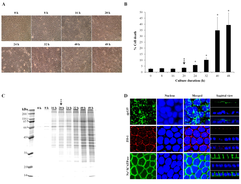
Figure 1. Optimal time-point for secretome analysis of polarized MDCK cells in serum-free medium.
Polarized MDCK cells were maintained in serum-free medium for various time-points, i.e. 0–48 h. Morphological examination was performed under a light microscope (A) (original magnification was 400X), whereas cell death analysis was done by using Trypan blue staining (B) (n = 3 independent experiments for each bar and *represents p < 0.05 as compared to 0 h). Proteins recovered from the culture supernatant of varying time-points (with an equal volume of 1 ml) were resolved by 12% SDS-PAGE and visualized with CBB-G250 staining (C). An arrow indicates the optimal time-point selected for secretome analysis (the longest duration that cell death remained unchanged – to ensure that secretome was analyzable whereas severe cytotoxicity could be excluded). Finally, immunofluorescence staining of apical (gp135), tight junction (ZO-1), and basolateral (Na+/K+-ATPase) markers was performed to confirm that the cells cultivated in serum-free medium for 20 h (optimal time-point) remained polarized and intact (D).

