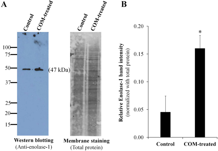
Figure 3. Validation of the proteomic data.
The increased level of secreted enolase-1 in COM-treated group determined by proteomic analysis was confirmed by Western blot analysis. Enolase-1 band was detected using rabbit polyclonal anti-enolase-1 antibody, whereas Deep Purple was used for membrane staining to normalize band intensity. N = 3 independent experiments; *p < 0.05 vs. control.

