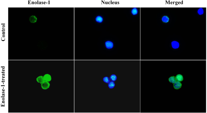
Figure 5. Binding of exogenous enolase-1 on U937 monocytic cell surface.
U937 monocytes were incubated with 500 pM purified enolase-1 for 24 h and then washed with PBS. Surface expression of enolase-1 was then examined by immunofluorescence staining (without permeabilization) using rabbit polyclonal anti-enolase-1 as the primary antibody. Chicken anti-rabbit IgG antibody conjugated with AlexaFluor-488 (green) served as the secondary antibody, whereas Hoechst dye (blue) was used for nuclear staining. The images were captured under a fluorescent microscope (Original magnification power = 1,000X).

