Figure 5.
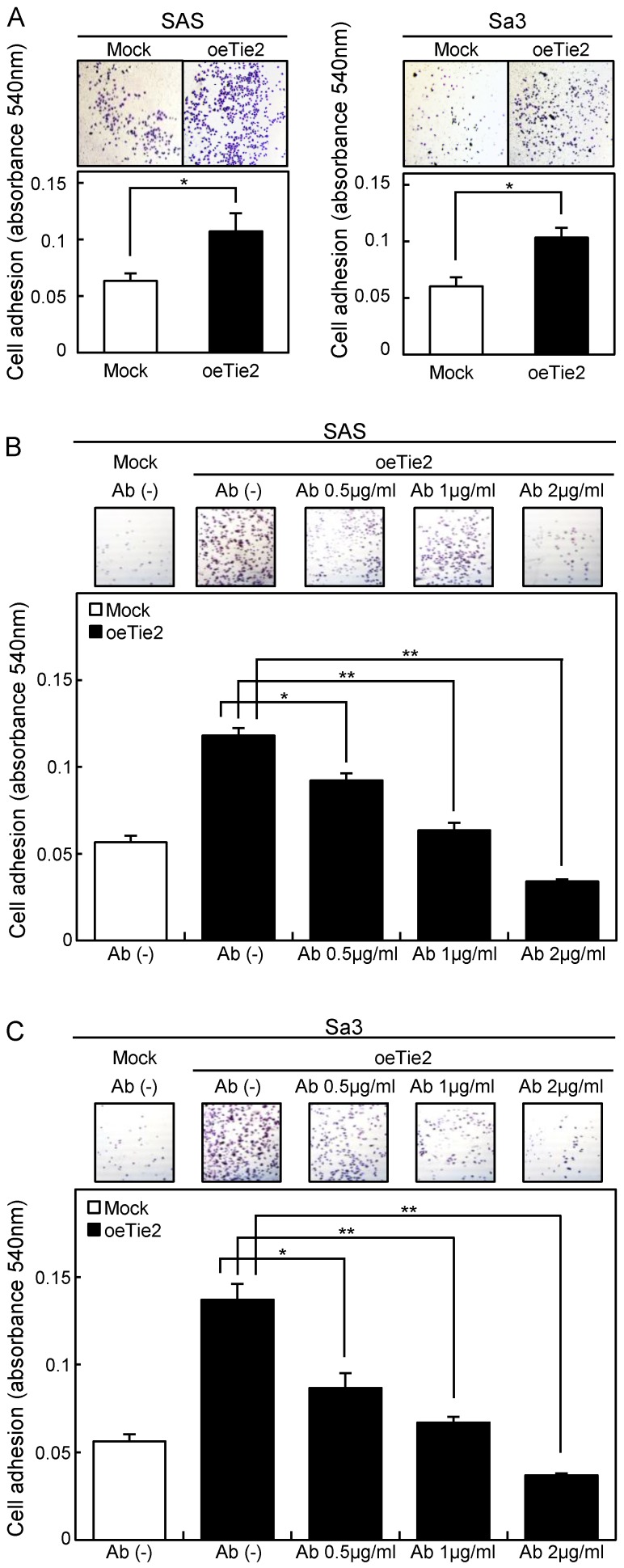
Adhesion assay of Tie2 overexpressed cells. (A) Adhesion assay of Tie2 overexpressed cells. To evaluate the adhesion ability, oeTie2 and Mock cells are seeded on collagen I-coated 96-well plates at a density of 2 ×10⁴ cells/well and allowed to adhere for 1 hour. After crystal violet staining, the numbers of stained cells are measured using a microplate spectrophotometer (absorbance at 540 nm and at 405 nm to subtract background). The cellular adhesion of the oeTie2 cells increased significantly (p < 0.05) compared with the Mock cells. (B, C) Inhibition of cellular adhesion by Tie2 antibody. oeTie2 cells are treated with Tie2 antibody before the adhesion assay. The numbers of oeTie2 cells attached to the dishes are decreased in a dose-dependent manner of the Tie2 antibody (B, SAS cells; C, Sa3 cells).

