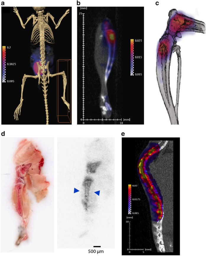
Figure 2.
Localization of [18F]-FDG in bone. (a) Whole-body PET/CT image of 6-week-old C57BL/6 male mouse 2 h after [18F]-FDG administration. (b) [18F]-FDG uptake in the right hind limb of the mouse from a. (c) Computer rendering of PET/CT image of [18F]-FDG uptake of the distal femur and proximal tibia of mouse from a. (d) Whole-mount tissue section of 6-week-old C57BL/6 male mouse hind limb with autoradiograph on the right showing [18F]-FDG uptake in the distal femur and proximal tibia. Arrows denote uptake in the cortical bone compartment. (e) PET/CT image of the spinal column of a 6-week-old C57BL/6 male mouse. Images are representative of five mice. Intensity thresholds map counts per second per voxel.

