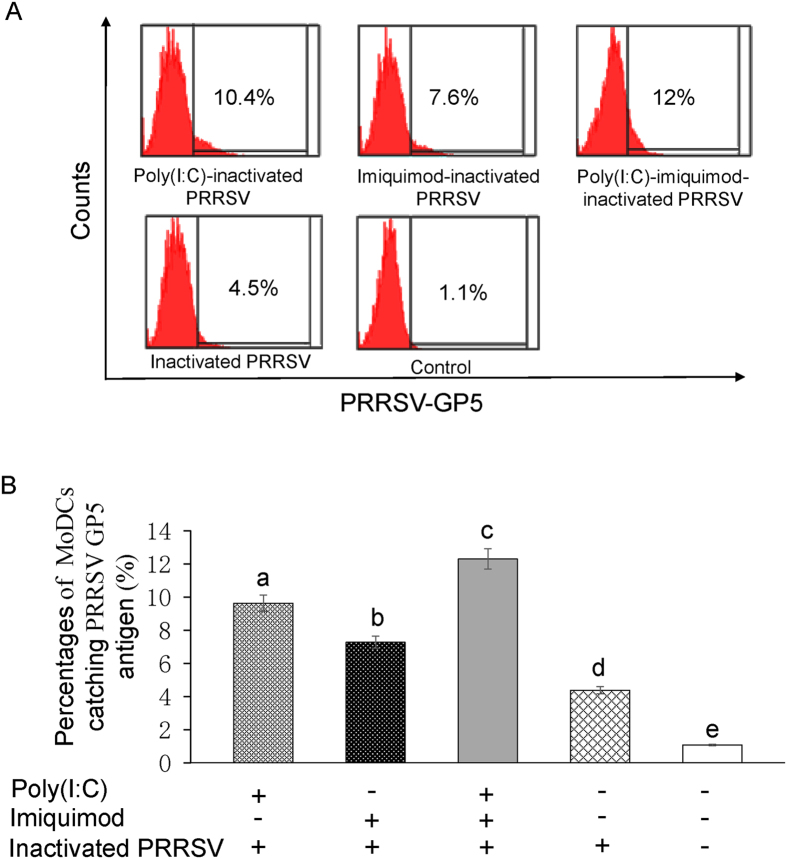
Figure 5. Phagocytosis of MoDCs treated with TLR3 and 7 ligands along with inactivated PRRSV antigen.
MoDCs were incubated with poly (I: C) and/or imiquimod along with inactivated PRRSV antigen for 12 h. MoDCs were fixed and permeabilized and then stained with monoclonal antibody against PRRSV GP5. After washing with PBS, cells were incubated with Alexa Fluor 488-conjugated goat anti-mouse IgG(H + L) for flow cytometry. (A) Representative flow cytometry profile of the percentages of MoDCs catching PRRSV GP5 antigen. The data presented here are results from one experiment of three flow cytometry experiments. (B) The statistical graph of the percentages of MoDCs catching PRRSV GP5 antigen. The data were analyzed using Flowjo7.6 software. Data represent the means ± standard deviations (error bars) of three independent experiments. Different letters (a–e) mean significant difference (P < 0.05).

