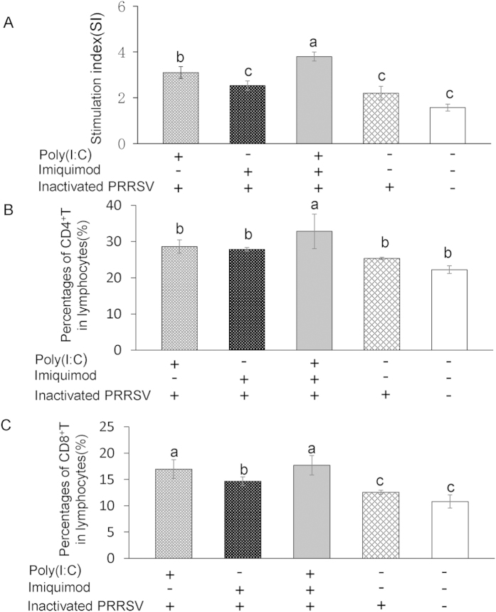
Figure 6. Proliferation of PRRSV-specific T lymphocytes and percentages of CD4+, CD8+ T lymphocytes in immunized mice.
Splenic lymphocytes were isolated from the spleen of vaccinated mice at 49 dpi. Part of splenic lymphocytes was stimulated with purified inactivated PRRSV antigen. Following 72 h incubation, the proliferation response was detected by a standard MTT assay. The PHA control sample showed a stimulation index of 5–7 (A). Another part of splenic lymphocytes was subjected to flow cytometry to assess the percentages of CD4+ T lymphocytes (B) and CD8+ T lymphocytes (C). Data represent the means ± standard deviations (error bars) of three independent experiments. Different letters (a–c) mean significant difference (P < 0.05).

