Figure 3.
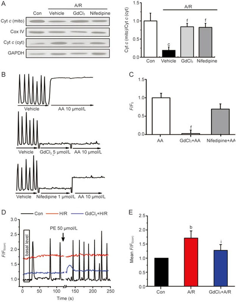
GdCl3 inhibited A/R-induced cardiomyocyte apoptosis via inhibition of the mitochondria-related signaling pathway. (A) Representative images of Western blots and quantitative analyses of cytochrome c in mitochondrial/cytosol fractions; (B) Typical traces represent the spontaneous Ca2+ transients in NRVMs before and after AA (10 μmol/L) treatment for 2 min in the presence or absence of GdCl3 (5 μmol/L) or nifedipine (1 μmol/L) for 2 min as indicated. (C) GdCl3 (5 μmol/L) inhibited AA-induced increase in [Ca2+]i in NRVMs. (D) Representative traces illustrate global Ca2+ transients before and after 50 μmol/L PE treatment in normal cells (Con) and the disability of spontaneous pacing in NRVMs with A/R or GdCl3+A/R treatment. (E) Basal [Ca2+]i levels, the mean fluorescence from 4 images at the beginning of recording normalized by the control F0, were significantly enhanced as a result of A/R, which was partially prevented by 5 μmol/L GdCl3. All values are presented as the mean±SD. n=3 for each bar in A. n=4–6 for each bar in B–E. bP<0.05, cP<0.01 vs control (A, E). fP<0.01 vs AA (C). iP<0.01 vs A/R.

