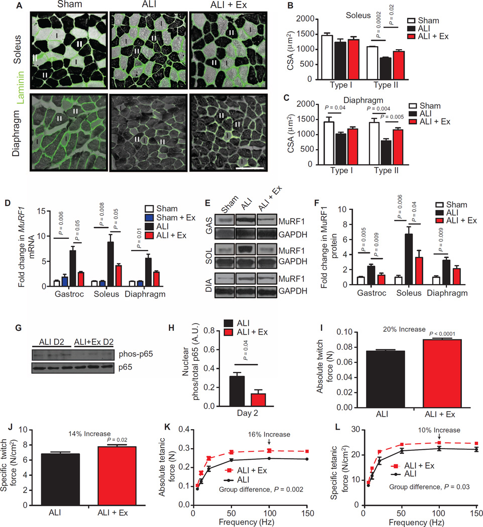
Fig. 3. Therapeutic exercise attenuates ALI-induced muscle atrophy and improves muscle performance.
(A) Type I (light) and II (dark) myofibers of the soleus and the diaphragm were identified by adenosine triphosphatase (ATPase) and laminin (green) staining. Scale bar, 100 µm. (B and C) Muscle fiber cross-sectional area (CSA) was quantified in sham, ALI, and ALI + Ex mice. (D to F) MuRF1 mRNA (D) and protein (E and F) levels normalized to GAPDH (glyceraldehyde-3-phosphate dehydrogenase) in gastrocnemius (GAS), diaphragm (DIA), and soleus (SOL) muscles. (G and H) Phosphorylated and total p65 protein from gastrocnemius nuclear extracts. D2 (day 2). (I to L) Ex vivo isolated soleus (I and J) absolute and specific twitch and (K and L) tetanic contractile force measurements. Values represent means ± SEM. All experimental time points are at day 3, other than (G) and (H), which are at day 2. n = 4 to 5 per group (A to D); n = 6 to 8 per group of two combined experiments (F); n = 3 per group (G and H); n = 4 animals and 8 muscles per group (I to L). Data were analyzed using the Student’s two-tailed t test or analysis of variance (ANOVA) for group differences with multiple time points.

