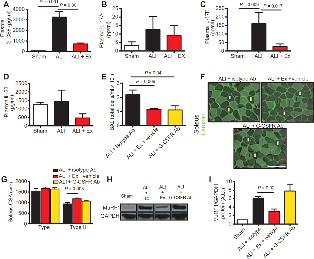
Fig. 5. Blockade of G-CSF activity limits lung injury but does not attenuate muscle atrophy.
(A to D) G-CSF, IL-17A, IL-17F, and IL-23 protein quantification in the plasma of sham, ALI, and ALI + Ex mice. (E) BAL cell counts were quantified at day 3 after systemic administration of isotype antibody (ALI + isotype Ab), exercise (ALI + Ex + vehicle), or G-CSFR–blocking antibody (ALI + G-CSFR Ab) 1 day after i.t.LPS administration. (F) Type I (light) and II (dark) myofibers of the soleus were identified by ATPase and laminin (green) staining. (G) Cross-sectional area was quantified in ALI + isotype Ab, ALI + Ex + vehicle, and ALI + G-CSFR Ab mice. Scale bar, 100 µm. (H and I) Soleus muscle lysates were probed for MuRF1 protein and normalized to GAPDH in sham, ALI + isotype Ab, ALI + Ex + vehicle, and ALI + G-CSFR Ab mice (H) and quantified by densitometry (I). n = 3 to 7 per group. Data were analyzed using the Student’s two-tailed t test.

