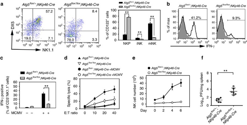
Figure 3. Autophagy is required for NK cell effector functions.
(a) NKP cells from Atg5flox/+;NKp46-Cre and Atg5flox/flox;NKp46-Cre mice were cultured in medium containing 10 ng ml−1 IL-15 on OP9 stroma cells for 7 days followed by cytometry. (b) CD3−NK1.1+ NK cells were isolated from Atg5flox/+;NKp46-Cre and Atg5flox/flox;NKp46-Cre mice and cultured in medium containing 10 ng ml−1 IL-12 and 10 ng ml−1 IL-18 for 12 h followed by brefeldin A (BFA) treatment, and stained for intracellular IFN-γ. Grey histograms depict isotype control of anti-IFN-γ antibody. (c) MCMV-induced IFN-γ is impaired in Atg5flox/flox;NKp46-Cre mice. Mice were infected with 1 × 10⁵ PFU MCMV for 3 days, and intracellular IFN-γ of NK1.1+ cells were tested by flow cytometry. Data were repeated for three times with similar results, and shown as means±s.d. **P<0.01. (d) Atg5flox/+;NKp46-Cre and Atg5flox/flox;NKp46-Cre mice were infected with MCMV for 3 days. NK cells were separated and incubated with 51Cr-labelled Yac1 cells with indicated E:T ratios. Specific lysis of Yac1 was analysed by 51Cr release, and shown as means±s.d. (e) Atg5flox/+;NKp46-Cre and Atg5flox/flox;NKp46-Cre mice were infected with MCMV for the indicated days, and NK cell numbers were analysed by flow cytometry. (f) Virus titers from MCMV-infected Atg5flox/+;NKp46-Cre mice and Atg5flox/flox;NKp46-Cre mice were analysed after three days' infection. n=6 mice for each group. **P<0.01. All data are representative of at least three independent experiments and calculated data are shown as means±s.d. **P<0.01. For a,c and f, a two-tailed unpaired Student's t-test was used.

