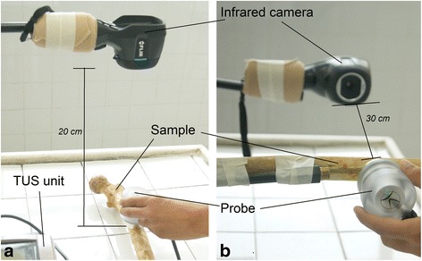
Fig. 2.

Experimental setups used in this study. a Experimental setup of the “surface heating” protocol showing the position of the infrared camera and TUS unit. Probe is above the sample (phantom or bone specimen). b Experimental setup of the “medullar canal” protocol. The infrared camera was positioned at 30 cm from the internal surface of the sample. The TUS probe was placed in contact with the sample on its external surface using coupling gel
