
Fig. 4.
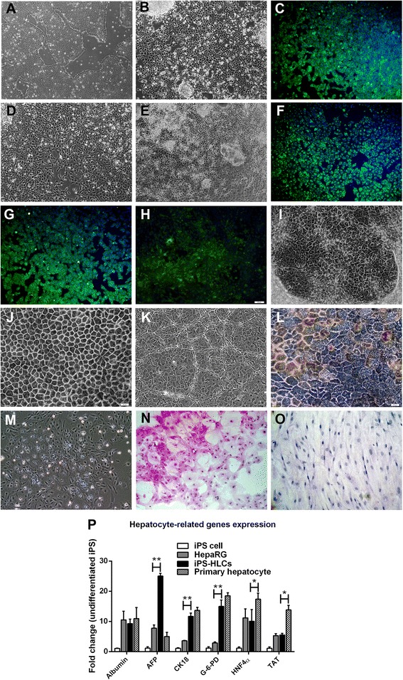
The direct differentiation of iPS cell into fully the functional homogenous HLCs. The iPS cells in feeder-free condition at 60 % confluence (a) were differentiated into definitive endoderm with round-shaped morphology (b). During differentiation induction, cells were positive for Sox17 (c). During anterior definitive endoderm (ADE) development, the differentiated cells underwent epithelial to mesenchymal transition (EMT) (d). The ADE cells with oval-shape (e) served as hepatic progenitors. Differentiated cells were positive for α-fetoprotein (f), albumin (g) and HNF-4α (h). The homogenously mature HLCs exhibited polygonal morphology (i), cord-like structure with bile canaliculi (j) and hepatic sinusoidal-like structures (k). The HLCs were positive for bile pigment (l). The 2nd passage of HLCs still maintained the hepatic morphology (m). HLCs and hMSCs were examined for glycogen storage using PAS assay (n, o). HLCs, HepaRG and primary human hepatocyte were compared for the expressions of hepatocyte selective genes (p). Data were presented as a fold changes over the undifferentiated iPS cells. * and ** represented statistical different data with a p value <0.05 or <0.01 respectively