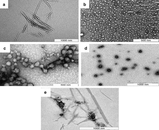
Fig. 1.

Transmission electron micrographs of DNPs: TEM image of a F∆F, showing the formation of tubular structure with mean diameter of 25 nm and length in microns, b M∆F, demonstrating the formation of vesicular structures with mean diameter of 40 nm c V∆F, showing the formation of vesicular structures with mean diameter of 55 nm, d RΔF demonstrating the formation of vesicular structures with mean diameter of 62 nm and e Ccm-F∆F showing dense tubular structures
