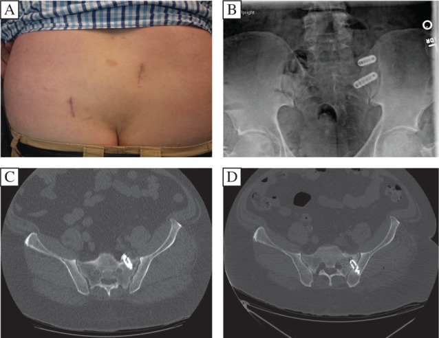
Fig. 4. Postoperative wound and imaging with revision.

A: Early postoperative wound appearance (reference arc wound, right; SIJ fusion wound, left). B: The post-operative x-ray demonstrates cage placement; however, the pre-revision X-ray was not helpful in confirming the proper cage position. C: Thus, a post-operative axial CT was performed. It demonstrated that the lower SIJ cage was placed deeply, breaching the ventral cortical wall of the sacrum and compressing the neural structure. D: An axial CT of the SIJ cage after revision demonstrated adequate positioning of the cage in the cortical bone.
