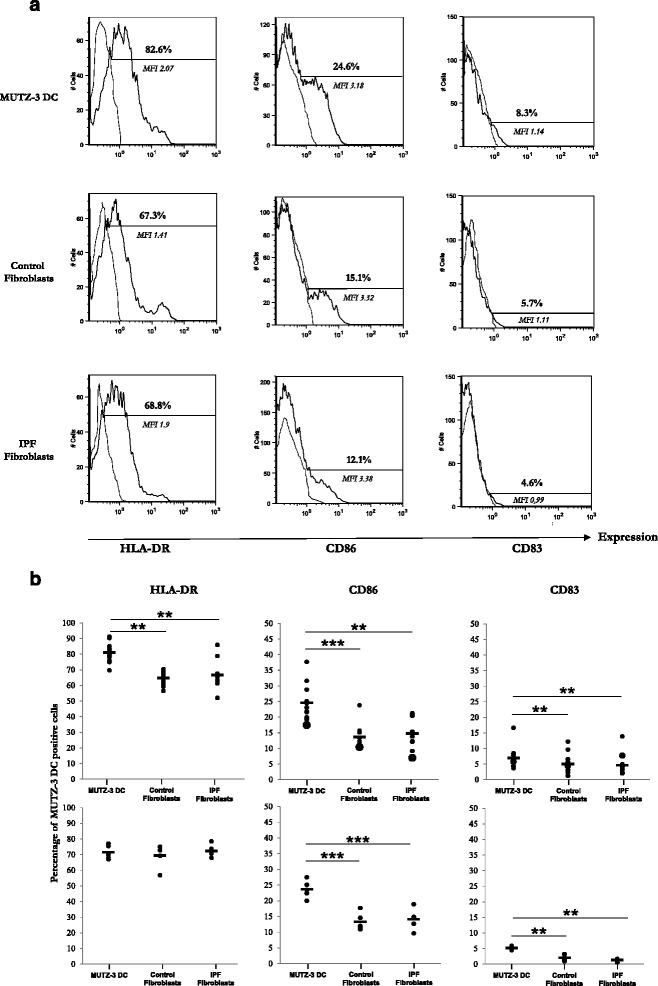
Fig. 1.

Phenotype of MUTZ-3 DCs alone or cultured with control or IPF fibroblasts for 48 h. Immature MUTZ-3 DCs (1×10⁶) were directly added to fibroblasts monolayers grown to confluence in 6-well tissue culture plates (direct contact) or fibroblasts (low compartment) and MUTZ-3 DCs (high compartment) were separated using modified Boyden chambers (Transwell® permeable support-0.4 μm) to prevent direct cell-cell contact, for 48 h in MUTZ-3 DC medium (Transwell condition). Controls were MUTZ-3 DCs cultured alone for 48 h in the same condition. Flow Cytometry analysis was assessed on DCs stained with monoclonal antibodies HLA-DR-PE (Pharmingen-BD), CD34-FITC, CD83-PE, CD86-PE, and CD209-PE (Immunotech-Beckman Coulter), and acquired with an Epics cytometer (Beckman Coulter). a One representative experiment or (b) percentage of positive cells for HLA-DR, CD86, and CD83 expression, represented as mean (dark horizontal bars) and individual values (n = 10) for direct contact co-cultures (upper panels) and Transwell® co-cultures (lower panels). MFI: Mean fluorescence intensity. **p < 0.01, ***p < 0.001. Statistical comparisons were performed with Wilcoxon paired nonparametric test for group comparisons or Kruskal-Wallis for unpaired nonparametric test, using Prism 5 (Graphpad Software Inc.)
