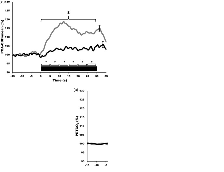
Figure 3.
Neurovascular coupling in humans generated from automated software using established standardized eyes-open/eyes-closed protocol. (a–c) Hyperemic responses, during visual stimulation, of the posterior cerebral artery (PCA; a), middle cerebral artery (MCA; b), and partial pressure end-tidal carbon dioxide (PETCO2; c). Note the selective activation of the PCA of around 20% vs. just 4−5% in MCA. Grey lines indicate healthy control group, black bars indicate high-level spinal cord injured group. Thick black bar indicates 30 s of eyes-open reading, being immediately preceded by eyes-closed. Smaller boxes represent 5 s bins which were averaged and compared quantitatively. These contours represent the response of 10 trials for each of 10 participants.

