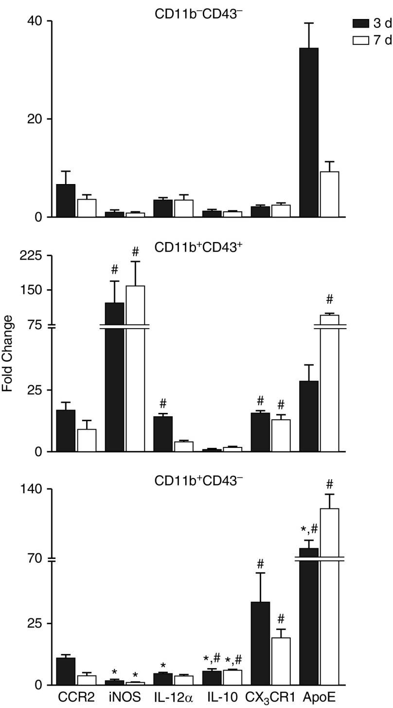
Figure 5.
Effects of NM on macrophage subpopulation gene expression. Macrophages, isolated 3 and 7 days after exposure of rats to PBS CTL or NM, were incubated with anti–rat-FcRII/III antibody, and then stained with antibodies to CD11b and CD43 as described in the Figure 4 legend. Viable cells (DAPI−) were sorted into CD11b−CD43−, CD11b+CD43+, and CD11b+CD43− subpopulations and processed immediately for RT-PCR analysis of expression of pro- (CCR2, iNOS, IL-12α) and anti- (IL-10, CX3CR1, ApoE) inflammatory genes. Data were normalized to glyceraldehyde 3-phosphate dehydrogenase and presented as fold change relative to resident CD11b−CD43− macrophages from CTL rats. Error bars, mean ± SE (n = 3–4 rats). *Significantly different (P ≤ 0.05) from CD11b+CD43+ macrophages. #Significantly different (P ≤ 0.05) from CD11b−CD43− macrophages.

