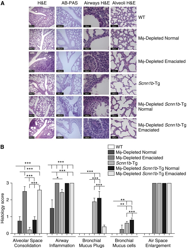
Figure 5.
MΦ-depleted neonates exhibit significant pulmonary pathology. (A) Representative micrographs for the genotypes, as indicated. Both emaciated and nonemaciated phenotypes are shown. Hematoxylin and eosin (H&E) stain was used to highlight morphological features, whereas Alcian blue/periodic acid–Schiff (AB-PAS) stain highlights mucus substances (dark purple). The scale bars in H&E (lungs), H&E (airways), H&E stained (alveoli), and AB-PAS stained (lungs) are 500 μm, 50 μm, 100 μm, and 200 μm, respectively. (B) Semiquantitative histological scores for relevant clinical features of neonatal mice. Data are expressed as means (±SEM). ANOVA: *P < 0.05, **P < 0.01, ***P < 0.001.

