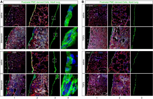
Figure 4.
Bleomycin induced infiltration with myofibroblasts but did not stimulate formation of mesenchymal cells from postnatal PMCs. Neonatal administration of tamoxifen labeled postnatal PMCs with GFP. Pulmonary fibrosis was then induced with bleomycin (A) or TGF-β1 (B). Infiltrating myofibroblasts strongly expressing vimentin or SMA did not coexpress GFP. Boxed areas are magnified in adjacent columns. In bleomycin treatment, the mesothelium became focally thickened (boxed areas, [A] column 3). Red lines ([A] column 4) indicate multiple layers of mesothelial cells. Focal mesothelial thickening was not observed with TGF-β1 treatment ([B] column 3). Scale bar = 100 μm.

