Figure 2.
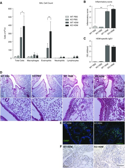
Wnt10b-deficient (Wnt10b−/−) mice exhibit increased inflammation compared with littermate WT mice after HDM challenge. (A) Bronchoalveolar lavage (BAL) cell counts show a significantly increased total number of cells and an increased number of eosinophils in Wnt10b−/− mice compared with littermate WT (n = 4–10/group, *P < 0.05 and **P < 0.01, respectively, t test; data compiled from two independent experiments, data are presented as mean ± SD). (B) Wnt10b−/− mice exhibit increased inflammatory infiltrates compared with their littermate controls after HDM challenge, shown by inflammatory index for severity of perivascular and peribronchial infiltrates (n = 3–4/group, 10 images/n; P < 0.05, t test). (C) No difference in sensitization measured by HDM-specific IgG1 was detected (n = 4–5/group, P < 0.05, t test). (D) Representative images of inflammatory infiltrates (upper panel scale bar, 250 μm; lower panel scale bar, 100 μm). (E) Immunofluorescent staining for eosinophil major basic protein also shows increased eosinophil infiltration in the lung parenchyma of Wnt10b−/− mice (representative images; scale bar, 100 μm). (F) Immunohistochemistry for CD3 does not show a clear difference in T cell infiltration in the lungs of Wnt10b−/− mice and controls (representative images; scale bar, 100 μm). OD, optical density.

