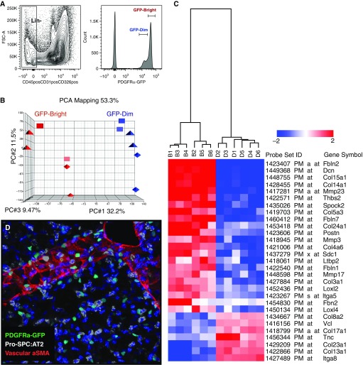
Figure 1.
Platelet-derived growth factor receptor α–green fluorescent protein (PDGFRα+-GFP) mice reveal two distinct fibroblast phenotypes. (A) Single cells isolated from adult mouse lungs were sorted into Lin⁻ (CD45⁻, CD31⁻, CD326⁻) and PDGFRα+GFPdim or GFPbright cells. (B) RNA was subjected to microarray analysis, and PCA mapping identified a myo- and a matrix messenger RNA signature. Using all probe sets, the first three principal components cover 75% of all variations across samples and clearly separate the two subpopulations (red and blue). (C) Collagen and matrix-related expression differences between PDGFRα-GFPbright and PDGFRα-GFPdim fibroblasts. Two-dimensional hierarchical clustering of 29 genes associated with extracellular matrix synthesis and collagen expression is differentially expressed between bright (B) and dim (D) cells. Red blocks represent up-regulation, and blue blocks represent down-regulated expression. (D) Three-dimensional confocal reconstruction of adult mouse lung demonstrate that PDGFRα-GFP+ cells (green) are adjacent to alveolar type-2 cells (pro-SPC; white) and are distributed evenly throughout the alveolar compartment. α-SMA, α-smooth muscle actin; FSC, forward scatter; GFP, green fluorescent protein; Lin−, lineage negative; PC, principal component; PCA, principal component analysis; pro-SPC, proprotein for surfactant protein C.

