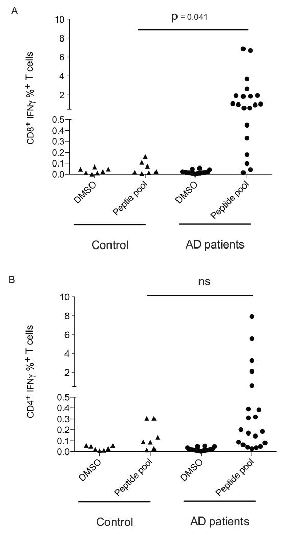
Figure 1. 21-OH specific T cells are detectable in Addison’s disease patients.
The production of IFN-γ from CD8+ (A) and CD4+ (B) T cells in response to 21-OH was assessed by culturing PBMCs from healthy controls or Addison’s patients for 14 days in the presence of a pool of peptides spanning the full length 21-OH protein, followed by stimulation for 12 hours with the 21-OH peptide pool (peptide pool) or with DMSO (DMSO). T cells were surface stained for CD4 and CD8 expression, then stained intracellularly for the production of IFN-γ. P-values were determined using an unpaired T test. Each dot corresponds to a different patient. Controls and pat ients have different symbols.

