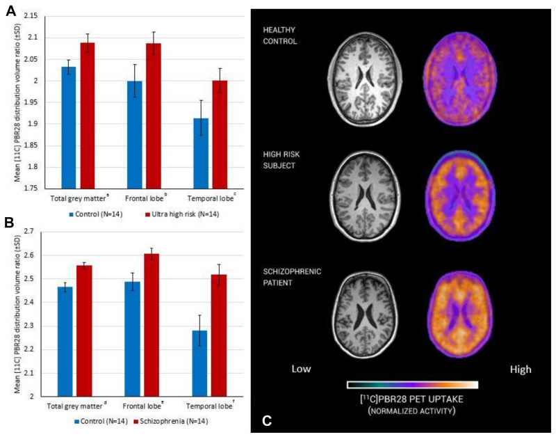
Figure 1. Microglial activity measured with PET in ultra high risk subjects, patients with schizophrenia and matched controls.
Significant difference between experimental (red) and control (blue) groups, ANCOVA (covarying for age and genotype). A: a (df=21 p=0.004), b (df=21 p=0.030), c (df=21 p=0.047); B: d (df=21 p<0.001), e (df=21 p=0.005), f (df=21 p=0.001); C representative [11C]PBR28 PET image from a subject from each group.

