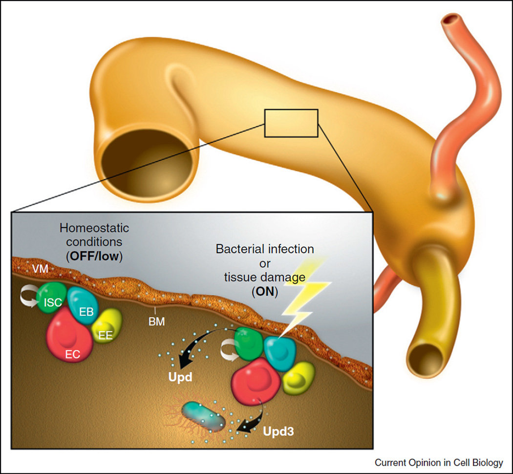
Figure 3.
JAK–STAT signaling in the posterior midgut. Schematic illustration of the midgut epithelium: Intestinal stem cells (ISCs, green), enteroblasts (EBs, blue), enteroendocrine cells (EEs, yellow), and enterocytes (ECs, red). Under homeostatic conditions, Upd is expressed in the adjacent visceral musculature and activates JAK–STAT signaling in ISCs and EBs. After tissue injury Upd cytokines are upregulated: Upd becomes expressed in progenitor cells (ISCs and EBs), while Upd3 is secreted by enterocytes.

