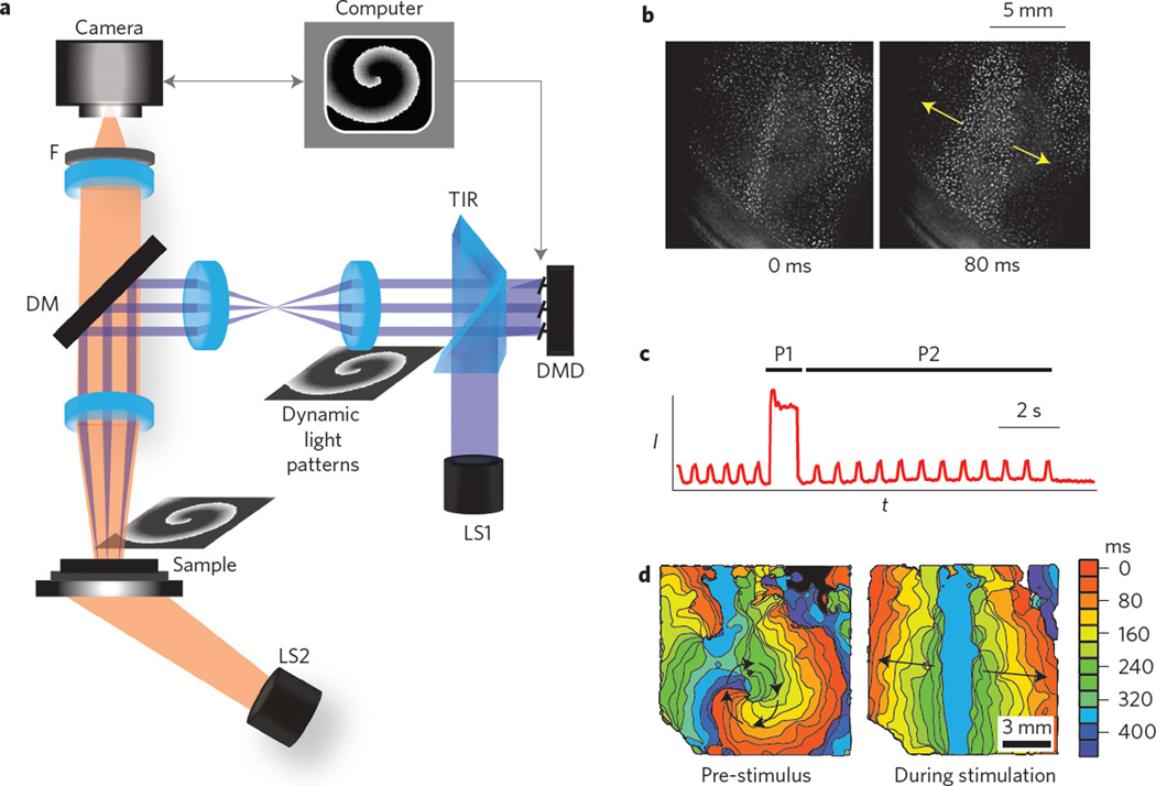
Figure 1. All-optical system for control of wave dynamics in biological media.
a, Experimental set-up, including an actuation light source (LS1: 10WLED, 460 nm), a total-internal-reflection prism (TIR) and a computer-controlled digital micromirror device (DMD). Generated light patterns are projected via lenses and a dichroic mirror (DM, 510 nm) to the biological sample. A second light source (LS2: white LED, bandpass filtered at 580 ± 20 nm) provides oblique trans-illumination for dye-free imaging onto a scientific complementary metal-oxide semiconductor (sCMOS) camera through an objective lens (×1, 0.25 NA) and a long-pass emission filter (F, >580 nm). b, Example of minimally filtered images in response to optical line stimulation in cardiac monolayers. c,d, Intensity (I) versus time (t) trace from a single pixel (c) and activation maps (d) showing ongoing spontaneous activity (a spiral) pre-stimulus, terminated by a strong global optical stimulation (P1) and followed by periodic optical stimulation (P2) by a line stimulus. For b–d, see also Supplementary Movies 1 and 2.

