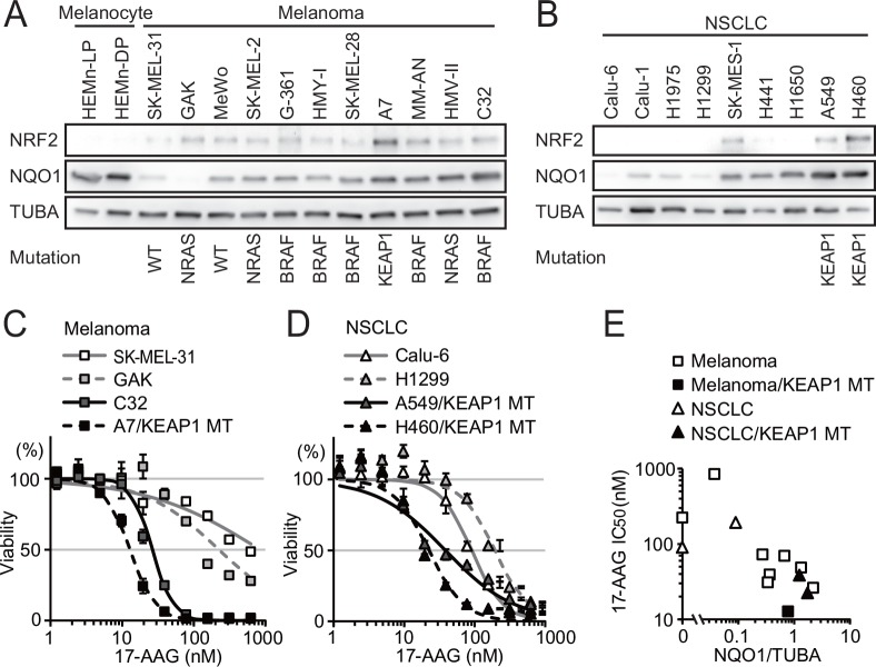
Fig 1. NQO1 expression and 17-AAG sensitivity in melanoma and NSCLC cell lines.
(A, B) Expression of NRF2, NQO1 and α-tubulin (TUBA) was detected by immunoblotting analysis in whole extracts of normal melanocytes, melanoma (A), and NSCLC cell lines (B). (C) 17-AAG sensitivity of melanoma cell lines with low NQO1 expression (SK-MEL-31 and GAK) and high NQO1 expression, with and without KEAP1 mutation (C32 and A7, respectively). (D) 17-AAG sensitivity of NSCLC harboring wild-type KEAP1 and NQO1-low (Calu-6 and H1299) and KEAP1-mutated NQO1-high cell lines (A549 and H460). (E) Relationship between NQO1 abundance and 17-AAG sensitivity of melanoma and NSCLC cell lines. The data were expressed as mean±S.D. of four independent experiments (n = 4).

