
Figure 3. Activation of Rac by Gαi2 requires p130Cas.
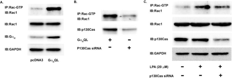
(A) Gαi2 activation is sufficient to lead to the activation of Rac1 in ovarian cancer cells. HeyA8 cells were transfected with either Gαi2QL, a constitutively active form of Gαi2, or pcDNA3 (vector). The cells were grown in normal media and not stimulated with exogenous LPA. After 48 hours, the cells were lysed and the lysates were immunoprecipitated with an antibody for the active form of Rac (Rac-GTP) overnight. The following day, the immunoprecipitated lysate was subjected to 10% SDS-PAGE and immunoblotted with an antibody for Rac1. 25 μg of lysate was subjected to 10% SDS-PAGE and immunoblotted for Gαi2 to demonstrate overexpression of the Gαi2QL vector. (B) Activation of Rac1 via Gαi2 requires the scaffold protein p130Cas. HeyA8 cells were transfected with Gαi2QL and with either non-targeting siRNA or siRNA directed against p130Cas as indicated. The cells were allowed to grow for 48 hours and the lysates were immunoprecipitated for active Rac for 24 hours. After 24 hours, the lysates were separated via 10% SDS-PAGE and immunoblotted for Rac1. To confirm knockdown of p130Cas, 25 μg of lysate was separated via 10% SDS-PAGE and immunoblotted for p130Cas. (C) p130Cas is required for Rac1 activation via LPA signaling. HeyA8 cells were transfected with either non-targeting siRNA or siRNA directed against p130Cas as indicated. After 24 hours, the transfected cells were serum-starved for 18 hours and then treated with 20 μM LPA for 20 minutes. The cells were then lysed and the lysates were immunoprecipitated with active Rac antibodies for 24 hours. The immunoprecipitated lysate was then subjected to 10% SDS-PAGE and immunoblotted with anti-Rac1 antibody. To confirm knockdown of p130Cas, 25 μg of lysate was subjected to 10% SDS-PAGE and immunoblotted for p130Cas. (D) p130Cas is localized to focal adhesions. HeyA8 cells were plated on glass slides and allowed to adhere overnight. The following day, the cells were serum-starved overnight and then stimulated with 20 μM of LPA for 5 to 60 minutes. The cells were then fixed and imaged for p130Cas and vinculin, a marker of focal adhesions. A representative image from 10 minutes is shown for p130Cas and vinculin co-localization and. (E.) p130Cas localizes to focal adhesions in a complex with activated Rac. Using the same treatment as Figure 3D, it was found that p130Cas and activated rac were localized to focal adhesions when cells were treated with 20 μM of LPA. Thus, it appears that p130Cas is localized mainly to focal adhesions and a sub-population of activated Rac is co-localized and directly interacting with p130Cas in the focal adhesions.