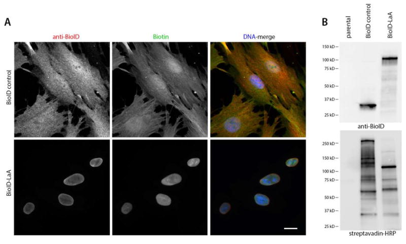
Figure 2. Proximity dependent biotinylation by BioID-LaA.

Control (non-transfected) human diploid foreskin fibroblast cells (BJ) or those cells stably expressing BioID-only or BioID-LaA (myc-BioID-LaA) by retroviral transduction were analyzed 18 hours after incubation in 50 μM biotin. (A) By IF analysis the BioID-only localizes to the nucleus and cytoplasm, whereas BioID-LaA (both red) localizes primarily to the nuclear envelope and minimally within the nucleoplasm. Biotinylated proteins, detected with labeled streptavidin (green), co-localize with the fusion proteins (red). DNA is labeled with Hoechst (blue). Bar, 10 μm. (B) IB analysis reveals migration of the fusion protein with anti-BioID and biotinylated endogenous proteins via streptavidin-conjugated HRP.
