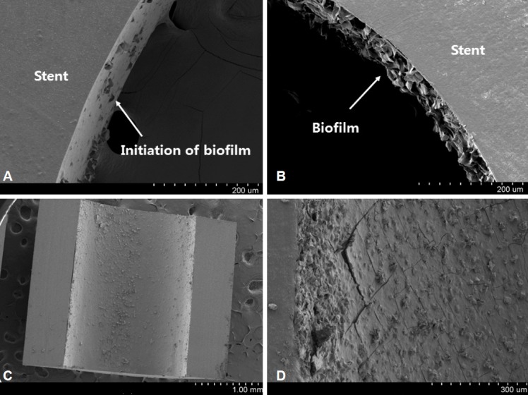
Fig. 2.

Scanning electron microscopy (SEM) examination of stent occlusion. SEM images of the inner surface of a stent retrieved at about 4 weeks. The biofilm starts to appear on the inner surface of the stent (A, ×250). The biofilm itself becomes gradually thicker relatively evenly (B, ×250; C, ×30). The surface becomes more solid and cracked in some areas (D, ×150).
