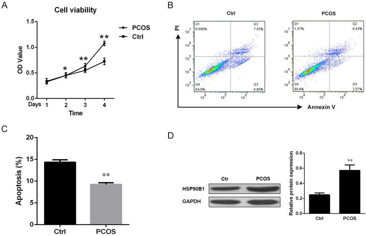
Fig 4. Cell apoptosis was decreased, but HSP90B1 was increased, in ovarian tissues from patients with PCOS.
(A) Cell viability was analyzed by the MTT assay. Purified ovarian cells from Ctrl and PCOS were cultured in RPMI1640 medium for the indicated durations. Data are presented as the mean of the OD value from triplicate wells ± standard error. One of three independent results is shown. (B) The cell suspension from ovarian biopsy was stained with propidium iodide (PI) and Annexin V conjugated to FITC. The stained cells were analyzed by flow cytometry, and one representative result is shown. (C) Apoptotic cells (Annexin V+PI-) were quantitatively analyzed, and the data are presented as the mean percentage of apoptotic cells ± standard error, n = 10. (D) HSP90B1 protein expression levels were analyzed by Western blotting. The blots were incubated with anti-HSP90B1 antibody (dilution 1:1000), and one representative result is shown (left panel). The band density was quantitatively analyzed by densitometric analysis, and the data are presented as the mean ratio of the target protein relative to GAPDH ± standard error, n = 10 (right panel).

