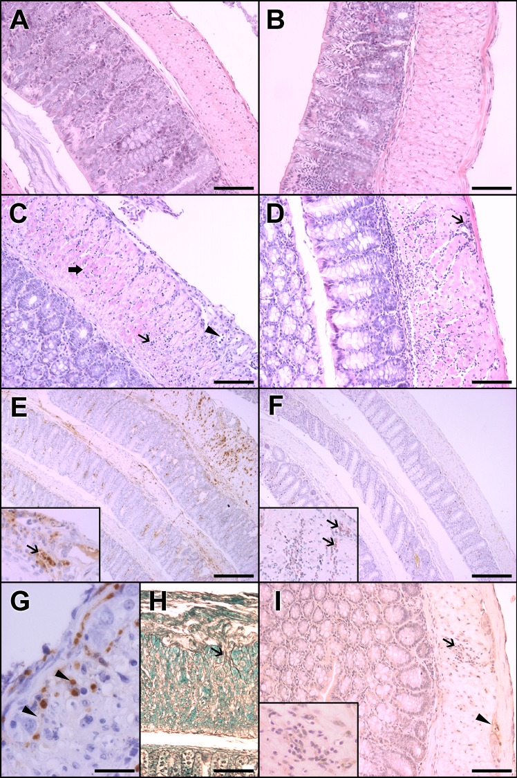
Fig 2. Histopathological features and evidence of parasitism in the colons of Swiss mice infected with 50,000 trypomastigotes of the Y strain of T. cruzi.
A, C, E, G, and H represent acute-phase aspects (11 d.a.i.). B, D, F, and I represent chronic-phase aspects (15 m.a.i.). (A) The colons of the control group of paired animals in the acute phase present normal cellularity and thickness. (B) In the chronic phase, no alterations were observed in the control group. (C) In acute-phase infected animals, a mononuclear inflammatory infiltrate was observed in the submucosal and muscular layers (arrow). In the myenteric plexus (arrowhead) and in the inner muscular layer, there is evidence of muscle fiber necrosis (thick arrow). This transmural pattern is merged with non-inflamed areas (not shown). (D) In contrast, in the chronic-phase infected group, mononuclear infiltrates are focal and more intense in the outer muscular layer in the periganglionar and perivascular areas (arrow). (E) Intense parasitism (inset,arrow) is associated with inflammatory infiltrates in the acute phase. (F) In the chronic phase, parasites are scarce and not associated with inflammatory foci (inset). (G) The acute-phase infected group showed strong iNOS positivity associated with inflammatory and degenerative changes in the myenteric plexus (arrowheads) compared (I) with the weak staining in inflammatory cells (arrow, and inset) in the chronic-phase infected group. A possible glial cell of ganglia (arrowhead) is also stained. (H) In addition, a greater amount of reticular fibers with thickened areas around the ganglia (arrow) were present in the acute phase. The results represent two independent experiments. A, B, C, D, I, H Magnification at 20x. Scale bar corresponds to 20 μm. E, F Magnification at 4x. Scale Bar corresponds to 100 μm. G Magnification at 40x. Scale bar corresponds to 10 μm. HE staining in A, B, C and D. Immunohistochemistry with anti-T. cruzi antibody in E, and F, and insets of E and F. Immunohistochemistry with anti-iNOS antibody in G and I. Silver Gomori staining in H.

