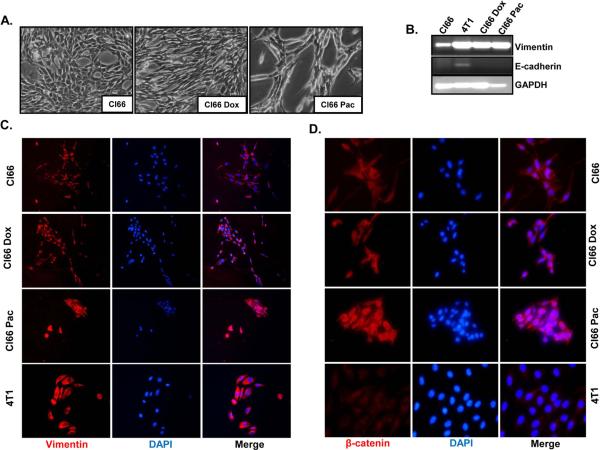
Figure 6. Drug-resistant Cl66 cells show transition from an epithelial to mesenchymal phenotype.
A) Photographic images showing in vitro cell morphology of parent Cl66, Cl66-Dox and Cl66-Pac cells. B) Expression of vimentin and E-cadherin in Cl66 parent and drug-resistant Cl66-Dox and Cl66-Pac cells by RT-PCR. Vimentin (C) and (D) β-catenin protein expression in parent Cl66, Cl66-Dox and Cl66-Pac cells examined by immunofluorescence. Blue color represents DAPI whereas red represents cy3 for vimentin (C) and β-catenin (D).

