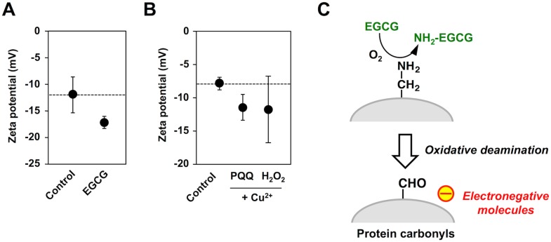
Fig 7. Formation of electrically-charged proteins by EGCG.
(A) Changes in the zeta potential of HSA treated with the catechins. HSA (1 mg/ml) was incubated with 1 mM catechins in 0.1 ml of PBS (pH 7.4) for 24 h at 37°C. (B) Changes in the zeta potential of BSA treated with the metal-catalyzed oxidation reactions. BSA (1 mg/ml) was incubated with 200 μM PQQ or 1 mM H2O2 in the presence and absence of 100 μM Cu2+ in 0.1 ml of PBS (pH 7.4) for 24 h at 37°C. (C) Schematic illustration of the EGCG-mediated transformation of HSA into electronegative molecules via oxidative deamination.

