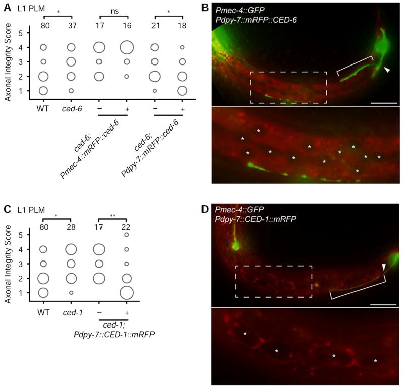
Figure 4. CED-1 and CED-6 function in the hypodermis.

(A) Cell-specific expression of Pmec-4∷mRFP∷CED-6 indicates that the molecule does not act cell-autonomously in PLM neuron, whereas expression of Pdpy-7∷mRFP∷CED-6 in the hypodermis is sufficient to rescue the diminished degeneration and clearance in ced-6(n1813) mutants. This strain is representative for three independent rescue strains (data not shown). (B) Localization of Pdpy-7∷mRFP∷CED-6 in the hypodermis in an animal at the L1 stage, 16 hours after axotomy. (C) Cell-specific expression of Pdpy-7∷CED-1∷mRFP is sufficient to rescue the diminished degeneration and clearance in ced-1(e1735) mutants. This strain is representative of three independent rescue strains (data not shown). (D) Localization of Pdpy-7∷CED-1∷mRFP in the hypodermis and seam cells in an animal at the L1 stage, 16 hours after axotomy. The area of each circle in panels A and C represents the proportion of data points that fall into that category; n value indicated on top of each bubble graph. * p<0.05, ** p<0.005, as determined by a Kruskal-Wallis and Dunn’s or a Mann-Whitney test. Scale bars indicate 20 μm. Arrowheads indicate the location of axotomy, brackets indicate regrowth, and asterisks mark the hypodermal and seam nuclei.
